Medical Imaging · 2026
Melanoma segmentation with U-Nets built from scratch in PyTorch
Two U-Nets coded from scratch to outline skin lesions: one trained from scratch and one with a frozen pretrained ResNet-50 encoder.
- pixel-level AUC-ROC on test
- 0.977
- IoU (Jaccard) on test
- 0.752
- IoU gain from the pretrained encoder
- +2.9 pts
- dermoscopic images with masks
- 2,750
Problem
Outlining the border of a skin lesion precisely is the first step to analysing it: asymmetry and border irregularity are classic signs of melanoma. The goal is a model that, given a dermoscopic image, predicts the lesion mask pixel by pixel (lesion or background).
The dataset is ISIC Melanoma Segmentation: 2,750 images with their annotated masks. The project’s question is whether it pays off to start from an encoder pretrained on ImageNet, a very different domain, instead of training the whole network from scratch.
My contribution
- I implemented a classic U-Net from scratch in PyTorch: five convolutional blocks with 64 to 1024 filters, a symmetric decoder with transposed convolutions and skip connections at every level (31 M parameters, all trainable).
- I implemented a U-Net with a frozen pretrained ResNet-50 encoder, with skip connections from
conv1,layer1,layer2andlayer3and a custom five-block decoder. It trains 26.5 M of its 50 M parameters. - I wrote the full pipeline: loading and preprocessing, training with early stopping, threshold selection and evaluation.
Technical approach
- Preprocessing512 × 512 px, greyscale, normalisation and binarised mask
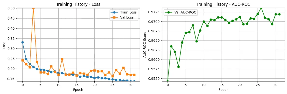
- TrainingAdam (1e-4) and BCE with logits, up to 50 epochsearly stopping: val. AUC
- Optimal thresholdPrecision-recall curve on validation; the max-F1 threshold is chosen
- Test evaluationPixel-level AUC-ROC and IoU (Jaccard index)
Images are converted to greyscale, so ResNet-50’s first layer, designed for RGB, does not fit. Instead of initialising it randomly, I averaged its weights across the three channels: it keeps what was learned about edges and textures and accepts a single channel.
# scripts/models/UNet_ResNet50_Encoder.py (excerpt)
resnet50 = models.resnet50(pretrained=True)
self.conv1 = nn.Conv2d(1, 64, kernel_size=7, stride=2, padding=3, bias=False)
# RGB to greyscale: average the weights of the three channels
self.conv1.weight.data = resnet50.conv1.weight.data.mean(dim=1, keepdim=True)

# Frozen encoder: only the decoder is trained
encoder = [self.conv1, self.bn1,
           self.layer1, self.layer2, self.layer3, self.layer4]
for layer in encoder:
    for param in layer.parameters():
        param.requires_grad = False
Two decisions in the evaluation protocol:
- Sampled AUC. Computing AUC over every validation pixel does not fit in memory; it is computed on one pixel in ten, which is enough to drive early stopping.
- Leakage-free threshold. The threshold that turns probabilities into a mask is chosen on validation (max F1 on the precision-recall curve) and then applied as-is on test. Choosing it on test would inflate the result.
Results
| Model | Trainable parameters | AUC-ROC (test) | IoU (test) | Threshold |
|---|---|---|---|---|
| U-Net from scratch | 31.0 M | 0.9616 | 0.7236 | 0.42 |
| U-Net with frozen ResNet-50 | 26.5 M | 0.9769 | 0.7524 | 0.34 |
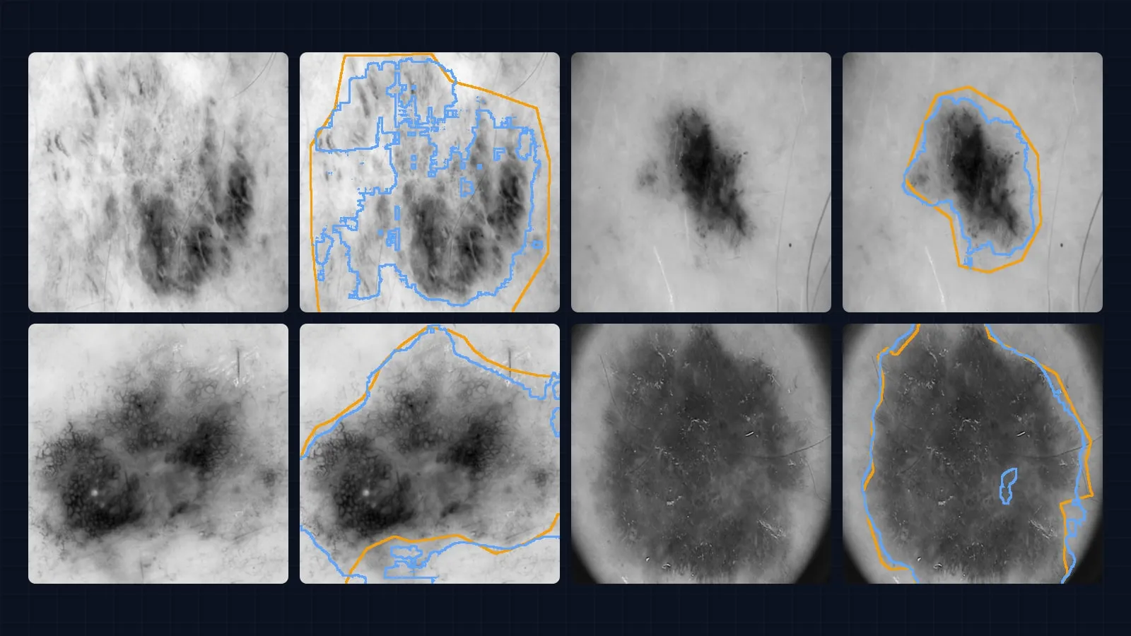
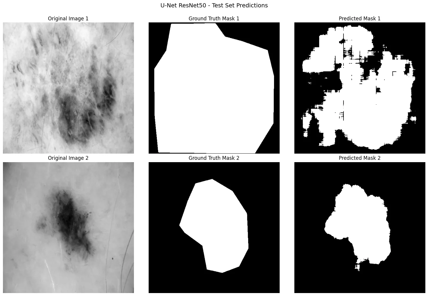
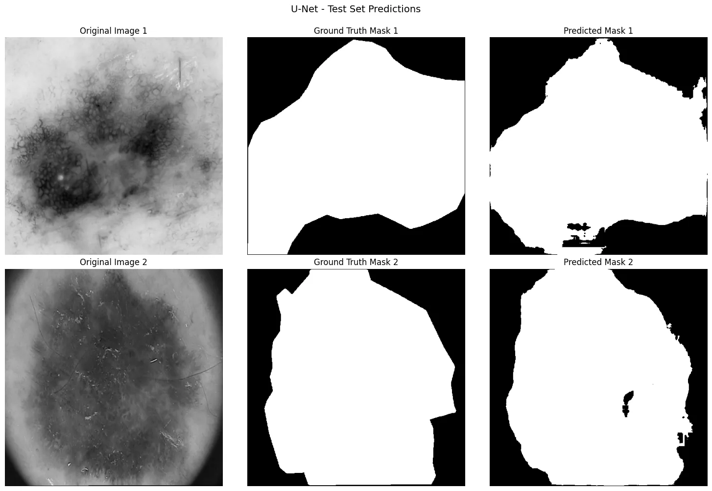
The pretrained encoder wins on both metrics (+1.5 AUC points and +2.9 IoU points) while training fewer parameters. ImageNet features, even from another domain, capture edges, textures and shapes that speed up convergence, and freezing them acts as regularisation with fewer than 2,000 training images.
 View full-size image
 View full-size image
 View full-size image
Evidence
- Repository with both architectures (
scripts/models/) and the training notebook with all its outputs.
Professional application
- Segmentation in medical imaging (lesions, organs, tumours) and other domains: defects in industrial inspection, plots in satellite imagery.
- Transfer learning with little labelled data: reuse a pretrained encoder and train only what is specific to the problem.
- Leakage-free evaluation protocols, with thresholds and model decisions always made outside the test set.
More projects
See all projects
Medical Imaging
Multi-label chest X-ray classification with DenseNet-121
Detecting 14 pathologies at once in 112,000 chest X-rays, with an asymmetric loss, per-pathology calibrated thresholds and Grad-CAM++ to audit the model.
Computer Vision
Ablation study on MNIST and CIFAR-10: 99.47% and 93.87% accuracy
Two ablation studies measuring how much each piece contributes to beating 99.4% on MNIST without convolutions and 92% on CIFAR-10.